”
As a biology enthusiast, I’m always fascinated by the intricate structures that make up living organisms. The animal cell, with its complex network of organelles and membranes, is a perfect example of nature’s remarkable engineering.
I’ve found that understanding animal cell structure is crucial for anyone interested in biology, medicine, or life sciences. Through detailed diagrams, we can explore the various components that work together to keep cells functioning – from the protective cell membrane to the energy-producing mitochondria. Whether you’re a student studying for an exam or simply curious about how living things work at their most basic level, having a clear grasp of cell anatomy will deepen your appreciation of biological systems.
Key Takeaways
- Animal cells are complex structures containing multiple organelles, each with specific functions and locations within the cell membrane
- The nucleus serves as the cell’s control center, housing DNA and regulating genetic material, while mitochondria generate energy through ATP production
- The cytoskeleton provides structural support and enables cell movement through three types of protein filaments: microfilaments, intermediate filaments, and microtubules
- Understanding cell diagrams requires familiarity with standardized labeling conventions, including line labels, leader lines, and color coding for different organelles
- Major cellular components can be identified by their distinctive visual characteristics – like the double-membraned nucleus, cristae-containing mitochondria, and interconnected endoplasmic reticulum
Biology:s3iownnwm48= Animal Cell Diagram
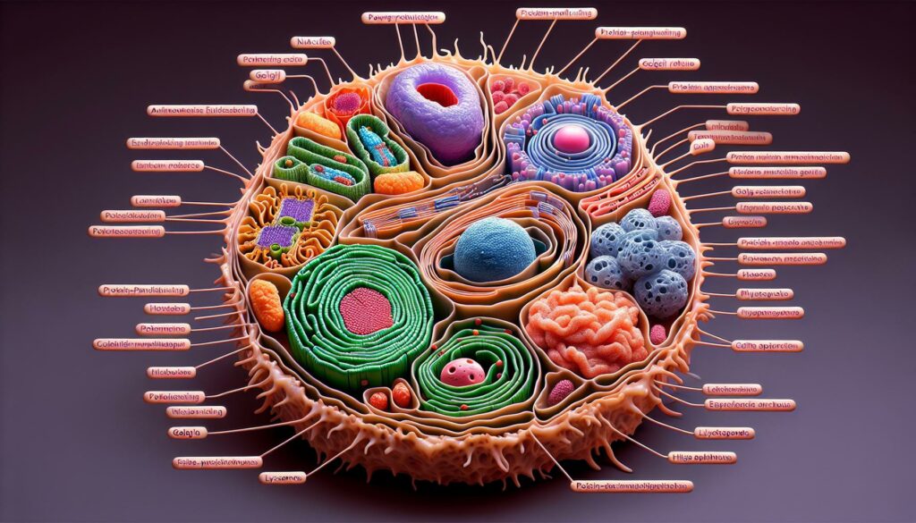
Animal cells contain distinct organelles arranged in a specific pattern within the cytoplasm. The organization of these components enables cells to perform essential life functions efficiently.
Key Components and Their Locations
Animal cells feature several essential organelles positioned strategically throughout the cell:
- Nucleus: Located centrally containing DNA genetic material
- Mitochondria: Scattered throughout cytoplasm generating cellular energy
- Endoplasmic Reticulum: Networks extending from nuclear membrane
- Golgi Apparatus: Positioned near nucleus for protein processing
- Lysosomes: Distributed in cytoplasm for waste breakdown
- Ribosomes: Found on rough ER surface or free in cytoplasm
- Centrosomes: Located near nucleus during cell division
- Vacuoles: Small structures dispersed in cytoplasm
| Organelle | Primary Function | Typical Size (μm) |
|---|---|---|
| Nucleus | DNA Storage | 3-10 |
| Mitochondria | Energy Production | 0.5-1 |
| Golgi Apparatus | Protein Processing | 0.5-1.5 |
| Lysosomes | Waste Digestion | 0.1-1.2 |
Cell Membrane and Cell Wall Differences
Animal cells possess a flexible phospholipid bilayer membrane without a cell wall:
- Membrane Components
- Phospholipids form fluid mosaic structure
- Embedded proteins control substance transport
- Cholesterol maintains membrane stability
- Glycoproteins enable cell recognition
- Structural Characteristics
- Flexible membrane allows shape changes
- Selective permeability controls molecular movement
- Direct cell-to-cell communication through membrane proteins
Essential Organelles in Animal Cells
Animal cells contain specialized structures that perform distinct functions to maintain cellular processes. Here’s a detailed examination of the primary organelles essential for cell survival.
Nucleus and Nuclear Membrane
The nucleus serves as the control center of the animal cell, housing genetic material in the form of DNA. The nuclear membrane, consisting of two phospholipid bilayers with nuclear pores, regulates the movement of molecules between the nucleus and cytoplasm.
| Nuclear Components | Key Functions |
|---|---|
| Nuclear envelope | Controls molecular transport |
| Nucleolus | Produces ribosomes |
| Chromatin | Stores genetic information |
| Nuclear pores | Facilitates selective transport |
Mitochondria and Energy Production
Mitochondria generate cellular energy through ATP production via cellular respiration. These double-membrane organelles contain inner folds called cristae that increase surface area for energy production.
| Mitochondrial Process | ATP Production |
|---|---|
| Glycolysis | 2 ATP |
| Krebs Cycle | 2 ATP |
| Electron Transport | 34 ATP |
Endoplasmic Reticulum and Golgi Apparatus
The endoplasmic reticulum (ER) exists in two forms: rough ER with attached ribosomes for protein synthesis and smooth ER for lipid production. The Golgi apparatus modifies, packages and distributes cellular products.
| Organelle | Primary Functions |
|---|---|
| Rough ER | Protein synthesis and transport |
| Smooth ER | Lipid synthesis and detoxification |
| Golgi apparatus | Protein modification and sorting |
| Transport vesicles | Product distribution |
Specialized Structures in Animal Cells
Biology:s3iownnwm48= Animal Cell Diagram contain specialized structures that enable movement mobility protection. I’ll examine two crucial systems that give cells their unique capabilities to maintain shape transport materials.
Cytoskeleton and Cell Movement
The cytoskeleton forms an intricate network of protein filaments throughout the cell. Three main types of filaments create this dynamic framework:
- Microfilaments (7nm diameter) create contractile forces for cell division movement
- Intermediate filaments (10nm diameter) provide mechanical strength structural support
- Microtubules (25nm diameter) form tracks for organelle transport cell division
The cytoskeleton enables cells to:
- Change shape during migration
- Maintain internal organization
- Create cellular extensions like cilia flagella
- Form the mitotic spindle during cell division
Vesicles and Transport Systems
Vesicles function as cellular cargo carriers moving materials between organelles. Key transport mechanisms include:
Transport Type
| Function |
Example Materials
|—|
Endocytosis
| Brings materials into cell |
Proteins nutrients
Exocytosis
| Releases materials from cell |
Hormones waste products
Transcytosis
| Moves materials across cell |
Antibodies ions
- Clathrin-coated vesicles for receptor-mediated endocytosis
- COPI vesicles for retrograde Golgi transport
- COPII vesicles for ER-to-Golgi transport
- Secretory vesicles for protein hormone release
Functions of Animal Cell Components
Biology:s3iownnwm48= Animal Cell Diagram components perform specialized tasks that maintain cellular homeostasis through coordinated processes. Each organelle contributes to specific functions that keep cells alive and operational.
Energy Processing and Storage
Mitochondria generate cellular energy by converting glucose into ATP through oxidative phosphorylation. These powerhouses contain inner membrane cristae that increase surface area for ATP production, producing up to 36 ATP molecules per glucose molecule. The cell stores energy in various forms:
- Glycogen granules store glucose for quick energy release
- Lipid droplets contain concentrated energy in fat molecules
- Mitochondrial matrix houses enzymes for the citric acid cycle
- Electron transport chain components line cristae membranes
- ATP molecules provide immediate energy for cellular work
- Ribosomes on rough ER synthesize proteins
- Transport vesicles move proteins from ER to Golgi
- Golgi apparatus modifies proteins through glycosylation
- Secretory vesicles package finished proteins
- Lysosomes contain enzymes for protein breakdown
- Signal sequences direct proteins to specific locations
| Organelle | Primary Function | Daily Production Rate |
|---|---|---|
| Ribosomes | Protein synthesis | 2 million proteins |
| Rough ER | Protein processing | 1000 different types |
| Golgi | Protein modification | 500,000 molecules |
| Lysosomes | Protein degradation | 50 different enzymes |
Reading and Interpreting Animal Cell Diagrams
Animal cell diagrams use standardized labeling methods to identify cellular components consistently across scientific literature. These visual representations follow specific conventions that enable accurate interpretation of cellular structures and their relationships.
Common Labeling Conventions
I identify three primary labeling methods in animal cell diagrams:
- Line labels connect specific structures to their names using straight or curved lines
- Leader lines point directly to smaller organelles with arrows
- Color coding distinguishes different organelles (e.g., red for nucleus, green for mitochondria)
Key formatting standards include:
- Capitalized labels for major structures (NUCLEUS, CYTOPLASM)
- Italicized labels for specialized components (e.g., nuclear pores)
- Numerical labels (1, 2, 3) with corresponding legends for complex diagrams
- Scale bars indicating actual cellular dimensions (typically 10-100 micrometers)
Notable Features and Identifiers
I recognize distinctive visual characteristics for major cellular components:
- Nucleus: Large circular structure with double membrane and visible nucleoli
- Mitochondria: Oval shapes with distinctive cristae formations
- Endoplasmic reticulum: Interconnected channels with attached ribosomes
- Golgi apparatus: Stacked membrane sacs with vesicles
- Lysosomes: Small circular structures with single membranes
- Cell membrane: Double-lined boundary with embedded proteins
- Membrane patterns (single vs. double)
- Internal structure details
- Relative size proportions
- Spatial relationships between organelles
- Specific shape characteristics
- Density variations in shading
Understanding Animal Cell Diagrams
Animal cells showcase nature’s remarkable engineering at its finest. From the protective membrane to the powerhouse mitochondria each component plays a vital role in sustaining life. I’ve explored how these microscopic structures work together in perfect harmony to keep organisms alive and thriving.
Understanding animal cell diagrams isn’t just about memorizing parts – it’s about appreciating the incredible complexity of life itself. I’m amazed at how these tiny cellular machines carry out countless tasks that make our existence possible. Whether you’re a student biology enthusiast or simply curious about life’s building blocks mastering animal cell structure opens the door to deeper insights into the fascinating world of biology.
“

